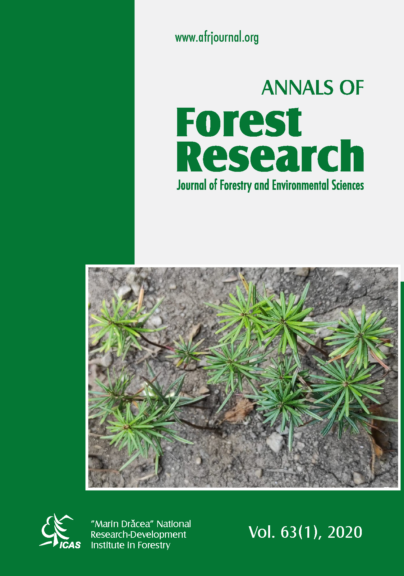
Micropropagation of an endangered species Pinus armandii var. Amamiana
DOI:
https://doi.org/10.15287/afr.2008.140Keywords:
micropropagation, Pinus armandii var. amamiana, conifer, somatic embryogenesisAbstract
For micropropagation via organ culture, mature embryos were excised from the seeds of Pinus armandii. Franch. var. amamiana (Koidz.) Hatusima, an endangered species only inhabiting the south west islands of Japan. Adventitious buds were induced on the surface of the embryo on 1/2 DCR medium containing BAP, and they grew shoots after subculturing to medium containing activated charcoal or a low concentration of thidiazuron. From the elongated shoots, root primordia and roots were induced in medium containing IBA as an auxine. We found that a low concentration of zeatin or BAP added to the medium was beneficial for plant regeneration of mature embryos of this species. For micropropagation via somatic embryogenesis, embryogenic cell suspensions were induced from a mature and immature seed of P. armandii var. amamiana on MS liquid medium supplemented with 1 ľM 2, 4-D and 3 ľM BAP. The suspensions were incubated in the dark at 250. Induced suspension cells were transferred to ammonium free MS liquid medium supplemented with 1 ľM 2, 4-D, 3 ľM BAP and 30m M L-glutamine and subcultured every 2 weeks. In the other set of the experiment, the induction rate of somatic embryogenesis was high with ammonium free half strength MS medium. In order to develop somatic embryos, the suspension cells were transferred to ammonium free MS medium supplemented with 10 ľM ABA, 0.2% activated charcoal, 10% PEG (MW6000), 30m M L-glutamine and 6% maltose. The cultures were incubated under a 16h light/8h dark photoperiod. After 1-2 months of culture, differentiation of embryos progressed and cotyledonary embryos were obtained. These embryos were transferred on ammonium free MS solid medium under 16 h photoperiod. After 2-3 weeks plantlets with roots and green cotyledons were obtained. Plantlets were transplanted to vermiculite containing modified MS liquid medium in 200 ml culture flasks, then out planted after habituation procedure.Downloads
Published
Issue
Section
License
All the papers published in Annals of Forest Research are available under an open access policy (Gratis Gold Open Access Licence), which guaranty the free (of taxes) and unlimited access, for anyone, to entire content of the all published articles. The users are free to "read, copy, distribute, print, search or refers to the full text of these articles", as long they mention the source.
The other materials (texts, images, graphical elements presented on the Website) are protected by copyright.
The journal exerts a permanent quality check, based on an established protocol for publishing the manuscripts. The potential article to be published are evaluated (peer-review) by members of the Editorial Board or other collaborators with competences on the paper topics. The publishing of manuscript is free of charge, all the costs being supported by Forest Research and Management Institute.
More details about Open Access:
Wikipedia: http://en.wikipedia.org/wiki/Open_access


